Top iridology camera, iriscope Suppliers and Manufacturer

FACEBAOOK | Youtube| Inlinked | X | IN

Iridology map: a practical clinic guide for independent wellness practitioners in Australia

» BLOG » Iridology map: a practical clinic guide for independent wellness practitioners in Australia

Iridology map: a practical clinic guide for independent wellness practitioners in Australia

November 2, 2026

Iridology map: a practical clinic guide for independent wellness practitioners in Australia — MAIKONG Iridology CHART (1).jpg
comparable provider map: a practical clinic guide for independent wellness practitioners in Australia · body

Quick answer

a comparable provider map is a reference chart that divides the iris into zones linked to anatomical regions and systems; in a consultation you use it as an observation tool to locate features (pigment, lines, rings, atrophy) relative to those zones. It’s a visual aide for recording and discussing findings with clients, not a diagnostic instrument. For Australian independent practitioners the useful decisions are: can a comparable provider map improve your consultation clarity and records, do you need dedicated imaging (high‑resolution iris camera and consistent lighting), and what professional limits and regulatory cautions apply. This article gives a clinic-ready workflow to match a client iris photo to a map, explains core zones and common markings, lists image-quality requirements and how MAIKONG cameras support them, and shows what to ask for when requesting equipment or chart materials.

Contents

What a comparable provider map is and how you can use it in a consultation

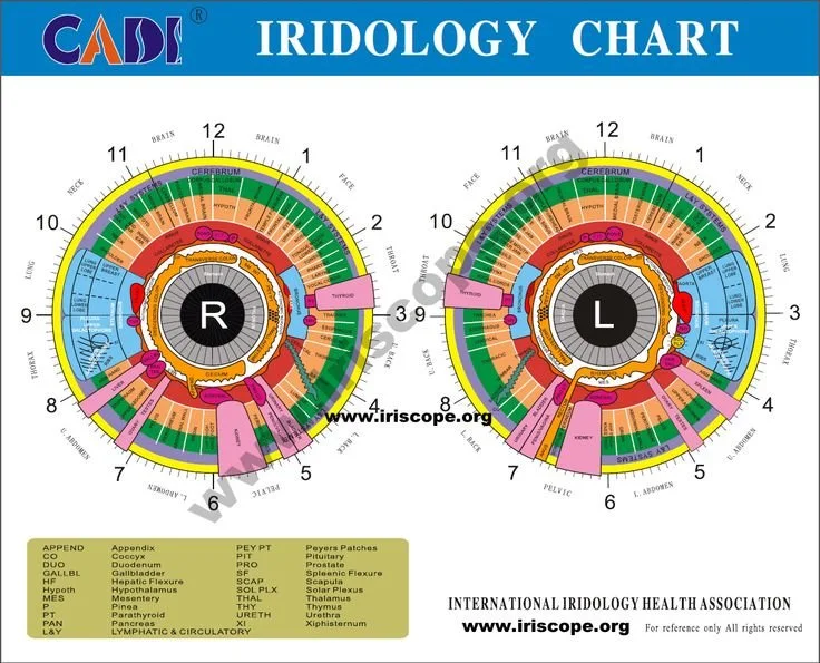
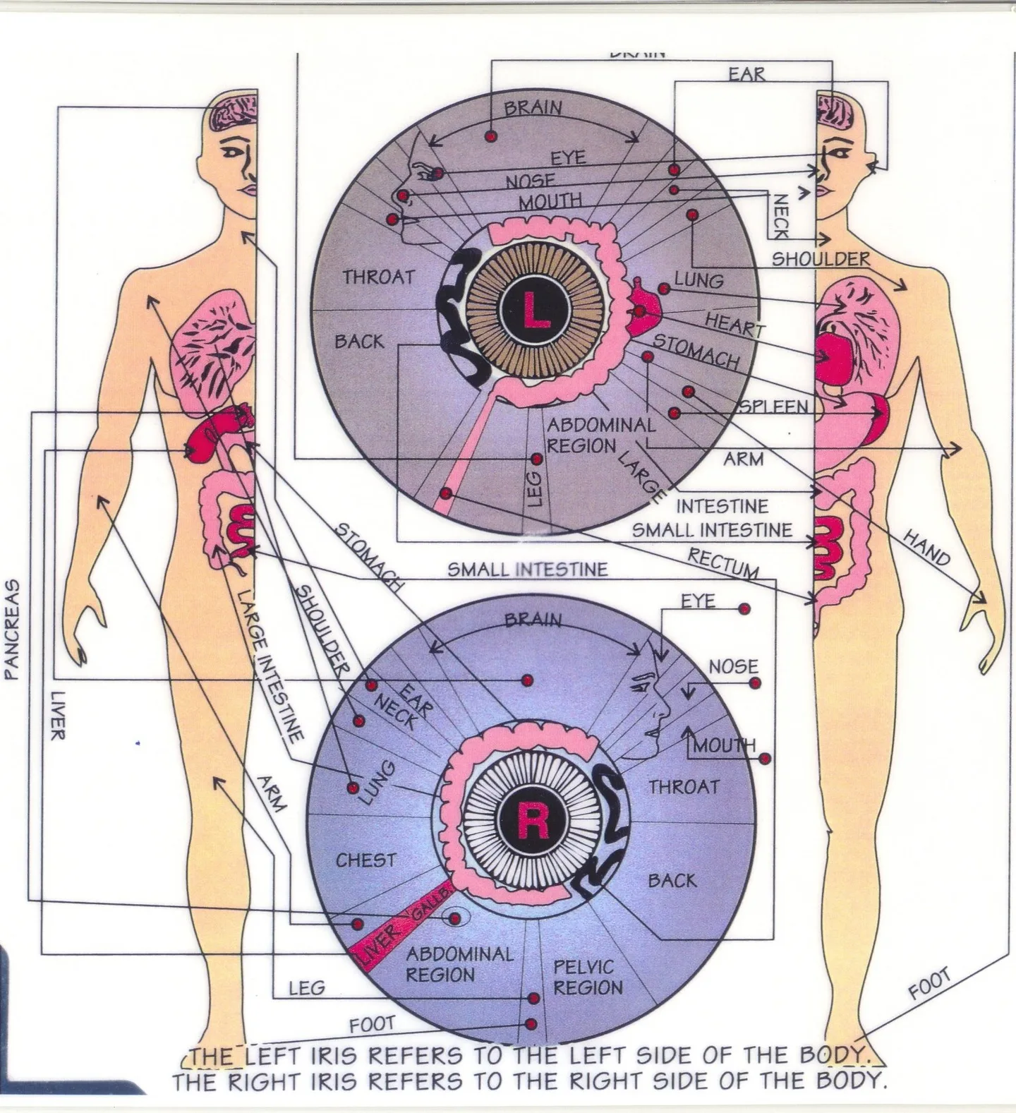
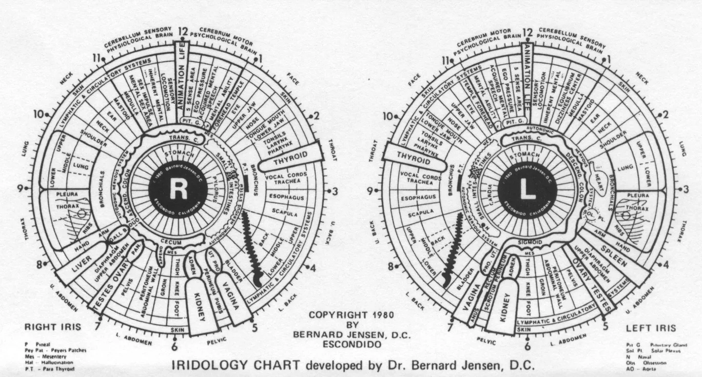
a comparable provider map (sometimes called an iris chart) overlays a standardised zone pattern on the iris so you can report where you observed a feature. In consultations we use it to:

  • locate and record client observations against a reproducible reference;
  • support client education by showing where a line, pigment spot or structural feature sits;
  • compare left and right irises and track changes between visits when images are archived.

The map is an observation framework — practitioners should not present map findings as medical diagnosis. Use it to structure notes, communicate visually, and guide further clinical enquiries or referrals.

Quick visual reference: how a typical comparable provider map aligns with the iris

Below is an explanatory overlay we commonly use in training and clinic reports. Use it to practise locating features on a photographed iris before you mark client records.
Explanatory iridology chart overlay on iris

The overlay divides the iris radially (clock positions) and concentrically (pupil zone, collarette, mid‑stroma, peripheral ring) so you and the client can say “2 o’clock, outer mid‑zone” rather than vague descriptions.

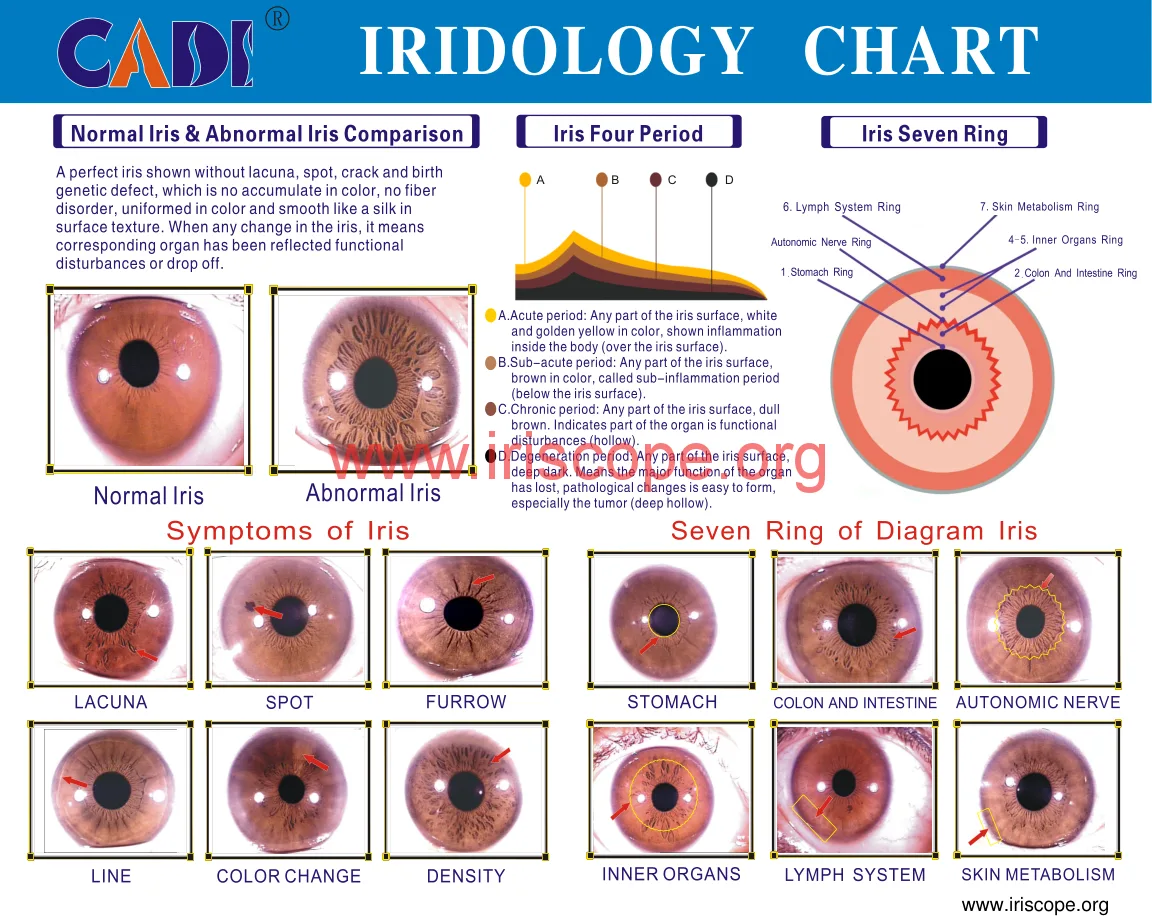
Core iris zones explained in clinic terms

Pupil zone

Closest to the pupil. Changes here (e.g. small central opacities) are recorded but often harder to capture because of eyelash reflection and pupil dynamics.

Collarette

The inner ring separating pupillary and sphincter fibres from the rest of the stroma. Collarette details (breaks, adhesions) are visual clues we note; describe them as present/absent and their clock position.

Radial furrows and spokes

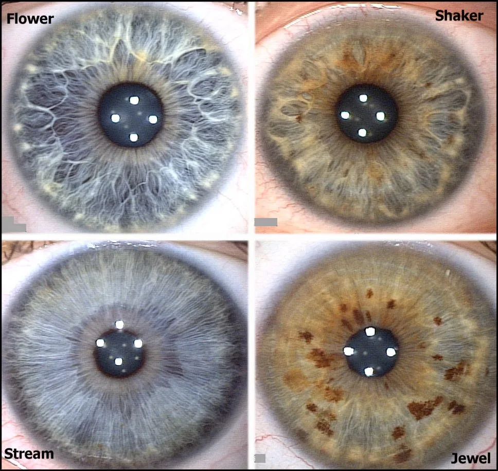
Lines that run outward. We record their density and whether they are single, branching or accompanied by pigment.

Nerve rings and contraction furrows

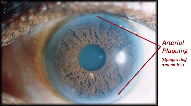
Concentric rings or tight wrinkling often visible near the outer stroma; useful as structural landmarks on the map.

Stroma and pigmentation areas

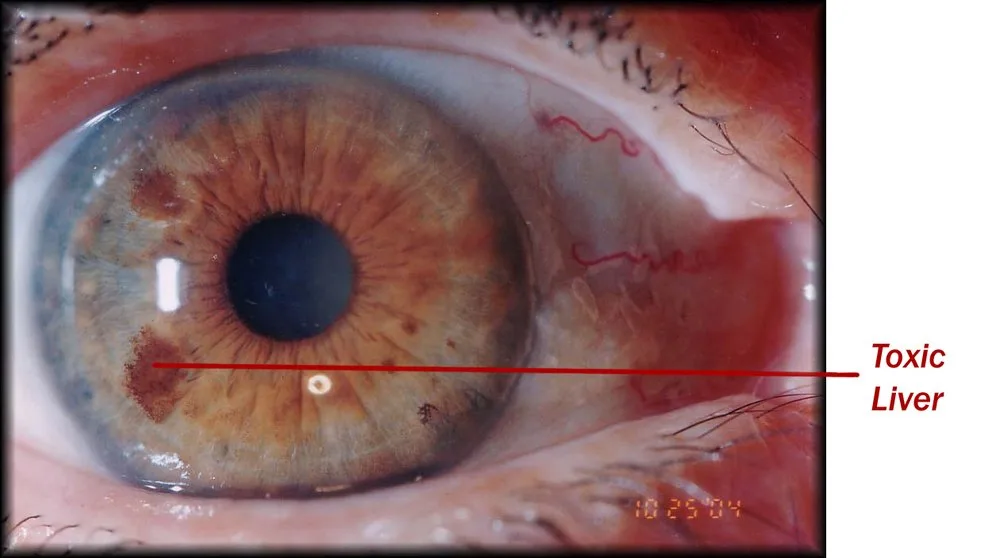
Colour variations, spots, or localised pigment clusters are documented by zone. Always note laterality (left/right) and record with an image.
Step‑by‑step: match a client iris photo to the comparable provider map

Iridology map: a practical clinic guide for independent wellness practitioners in Australia — MAIKONG Iridology CHART (2).png
comparable provider map: a practical clinic guide for independent wellness practitioners in Australia · body
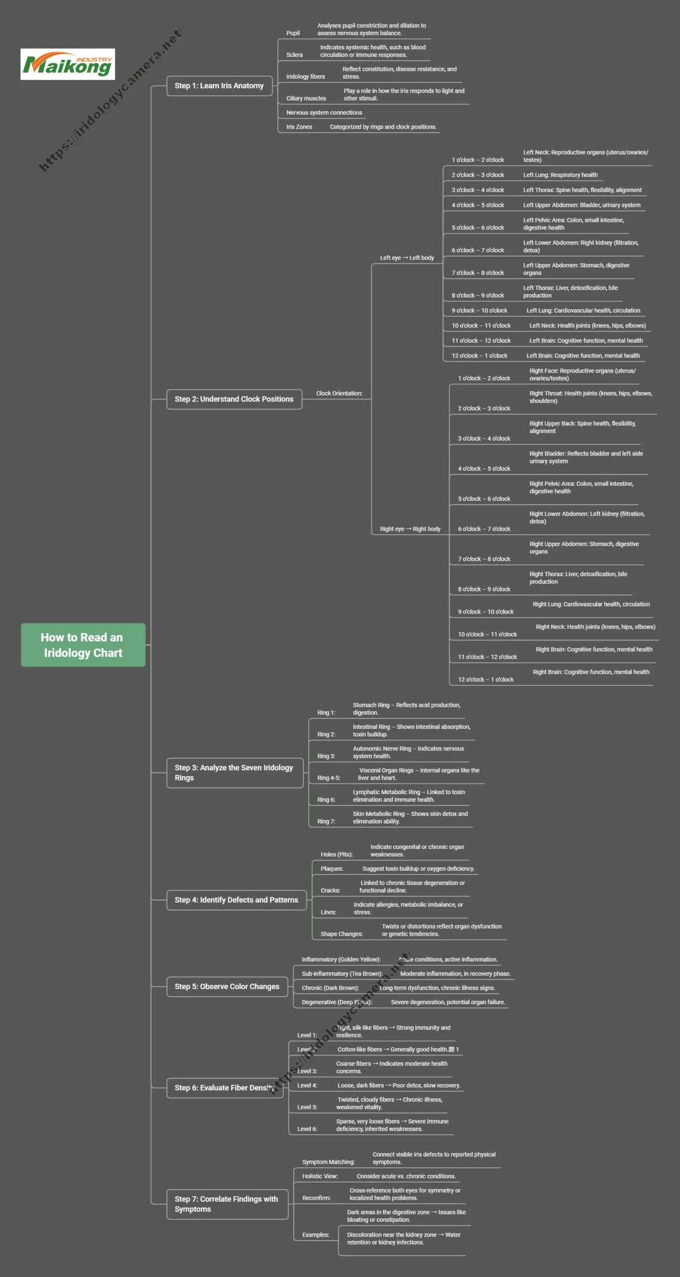
  1. Capture a high‑quality image (see Image quality checklist below).
  2. Open the image in your reporting software or on a printed chart and orient the photo so the 12 o’clock position is anatomically correct for that eye.
  3. Identify the pupil edge and the collarette — these are your concentric anchors.
  4. Overlay the chart or position a transparent chart print (300×420mm desk charts are common) and align using pupil/collarette; confirm radial markers match visible spokes.
  5. Mark observations as clock positions and concentric zone (pupillary, inner, mid, outer), and annotate whether each mark is a pigment, line, ring, or structural change.
  6. Save the annotated image into the client record with date and eye label (L/R).

If you use MAIKONG software, it supports image alignment and zone reference inside the application (see our comparable provider Software page for features and file support). For manual clinics, a printed desk chart and a transparency are often sufficient for teaching and record‑keeping (data-knowledge-id=”560959459″).
Using left/right comparison: when and why bilateral mapping matters

Iridology map: a practical clinic guide for independent wellness practitioners in Australia — MAIKONG Iridology CHART (10).jpg
comparable provider map: a practical clinic guide for independent wellness practitioners in Australia · body

Comparing left and right helps you spot asymmetry that may be clinically or educationally useful. In practice we:

  • always store both eyes under the same session record;
  • place images side‑by‑side and align pupil/collarette scale so clock positions match;
  • note whether a feature is unilateral or bilateral and whether it is fixed or variable between visits.

Bilateral mapping is not a diagnostic test; it’s a reproducible way to track observations and inform discussion or referral.

Image quality checklist: what a usable iris photo needs and how MAIKONG cameras support this

Iridology map: a practical clinic guide for independent wellness practitioners in Australia — MAIKONG Iridology CHART (8).jpg
comparable provider map: a practical clinic guide for independent wellness practitioners in Australia · body

For a clinic-quality comparable provider map match, every image should meet these minimal standards:

  • sharp focus on iris fibres and collarette;
  • even, controlled illumination (no hot spots across the iris);
  • visible pupil edge and collarette with minimal pupil constriction variation between captures;
  • resolution sufficient to show pigment spots and thin radial marks when image is viewed at consultation size.

MAIKONG camera details we reference when advising clinics:

Model Sensor / resolution Illumination OS / software

Common markings on the map and how to record them (observation, not diagnosis)

Iridology map: a practical clinic guide for independent wellness practitioners in Australia — MAIKONG Iridology CHART (14).jpg
comparable provider map: a practical clinic guide for independent wellness practitioners in Australia · body
  • Pigment spot — record clock position, size (small/medium/large), and permanence across visits.
  • Radial line/spoke — record start/end zones and whether single or branching.
  • Contraction furrow or nerve ring — note location and whether it interferes with reading other features.
  • Atrophic or thin areas — describe as localised stromal thinning and capture in high contrast lighting for clarity.

Use standard shorthand in your notes (e.g. “L eye 3 o’clock mid-zone: small pigment; unchanged vs last visit”). Keep observation language neutral and refer clinical concerns to a medical practitioner.
Limitations and professional cautions — what a comparable provider map can and cannot tell you in Australia

Iridology map: a practical clinic guide for independent wellness practitioners in Australia — MAIKONG iridology eye color (7).jpg
comparable provider map: a practical clinic guide for independent wellness practitioners in Australia · body

The map is an observational tool to structure and communicate findings. It cannot—and should not—be used as a substitute for medical assessment. We declare MAIKONG’s certifications (ISO 9001, CE, ISO 13485) as company statements; these relate to manufacturing and quality systems and are not endorsements that comparable provider is a diagnostic medical test. Australian practitioners must apply relevant advertising, consumer protection and health‑practice rules when describing services and equipment. Proper training in image capture and chart reading is essential before interpreting images for clients.
Practical tools and resources for clinics

Iridology map: a practical clinic guide for independent wellness practitioners in Australia — iridoligy (10).jpg
comparable provider map: a practical clinic guide for independent wellness practitioners in Australia · body

Items that make a comparable provider map usable day‑to‑day in a busy practice:

  • Universal comparable provider desk chart 300×420mm (useful as a printed reference in consult rooms; data-knowledge-id=”560959459″).
  • Flash cards for teaching or student clinics (useful for workshops; data-knowledge-id=”560959458″).
  • Report software that stores L/R images, overlays charts, and exports client PDF summaries (Eyeris‑style report modules and Maikong software options exist; data-knowledge-id=”560959590″).

If you plan to teach or run short courses, combine a printed chart, flash cards and software that lets students practice overlay alignment on saved images.
One‑page quick workflow you can print for consultations

Iridology map: a practical clinic guide for independent wellness practitioners in Australia — iridoligy (7).jpg
comparable provider map: a practical clinic guide for independent wellness practitioners in Australia · body

Iris capture → map alignment → annotate → save → discuss → refer if needed

  1. Seat client, neutral lighting, no strong makeup or reflective jewellery near eyes.
  2. Capture stable image; check focus on collarette and stroma. Save as YYYYMMDD_Lastname_L.jpg.
  3. Open image in software (or place beneath transparency). Align pupil/collarette to chart.
  4. Record features with clock notation, label L/R and date, attach image to client file.
  5. Use neutral language in notes; recommend further assessment for any health concerns.

Useful follow‑up questions to answer with further training or software

  • Do you need live overlay alignment in software or are printed charts sufficient for your workflow?
  • How will you store and back up client images (local server, cloud, encrypted drive)?
  • Do you want left/right comparison and change tracking across visits?
  • Which camera resolution fits your practice needs: 5MP for basic use, 12MP if you want larger archival crops and teaching detail?

Where to read more on our site

Practical next step

If you want a quote or configuration advice tailored to an Australian clinic (which camera resolution, software package and chart set best suit your workflow and teaching needs), email Lucy@iridologycamera.net with:

  • your intended use (consultation, teaching, demonstration),
  • which OS you run (Windows or Mac) and whether you need single or multi‑user licences,
  • quantity and destination (for shipping terms),
  • any required accessories (desk chart, flash cards, iris lenses).

Pricing and delivery details are provided on inquiry.

Images and product evidence

Representative device photography (for demonstration and installation reference):
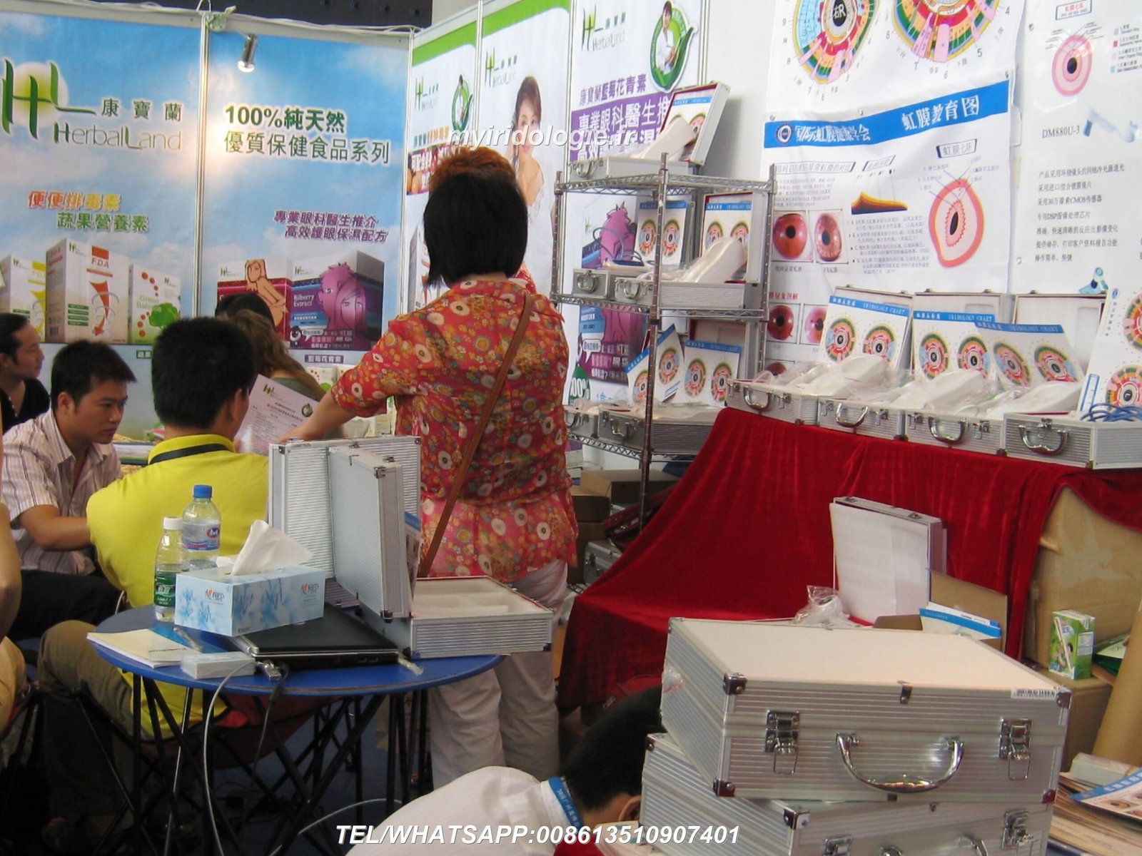
MAIKONG device exhibition photo

MAIKONG iridology camera on stand

Device specifications cited in this article come from MAIKONG product descriptions for MK 980U and MK-9822U (model identifiers and values preserved above); consult model pages for full accessory lists and software version notes.

FAQ (short)

Do I need a high‑resolution camera to use a comparable provider map?

Are MAIKONG cameras certified?

MAIKONG declares ISO 9001, CE and ISO 13485 certifications as company statements. These relate to production quality systems; they do not make clinical claims about comparable provider as a diagnostic method.

Compliance notice

This content is for educational and wellness purposes. MAIKONG iriscope, comparable provider camera, chart and software products support professional comparable provider education, image observation and record‑keeping. comparable provider is a complementary practice and not a medical diagnostic tool. Users should be appropriately trained. Product use and marketing in Australia must comply with applicable laws and professional practice requirements. Any health concerns should be referred to a qualified medical professional.

Contact

If you’d like a tailored configuration or quotation for your clinic, email Lucy@iridologycamera.net with the details requested in the “Practical next step” section.

This content is provided for learning, information sharing and professional communication only. It is not medical advice and is not a basis for treatment or diagnosis. Confirm suitability, operation and purchasing requirements against the product manual, local regulations and qualified professional guidance.

Discuss your requirements

Related resources

Discuss your requirements

Contact · WhatsApp · +8613510907401


Contact us online by Whatsapp:

marikongwhatsapp

Maybe you like also

Verified by MonsterInsights