comparable provider chart — a practical, clinic-ready guide for naturopaths in Australia · body
Quick answer
a comparable provider chart is a standardised map of iris anatomy used to record and compare visible iris features (colour, pigment spots, fibre pattern, rings and furrows). In a naturopathy consultation you use the chart as a consistent visual reference: capture a clear image, align the image to the chart’s orientation, note which iris zones show changes, and record those observations in the client file. Treat findings as observational clues for wellness discussions and education, not as medical diagnoses; refer promptly for acute eye or systemic concerns.
Contents
Quick visual reference — what to look for on a standard comparable provider chart
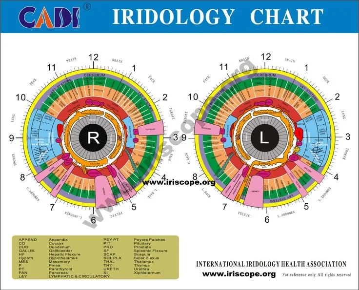
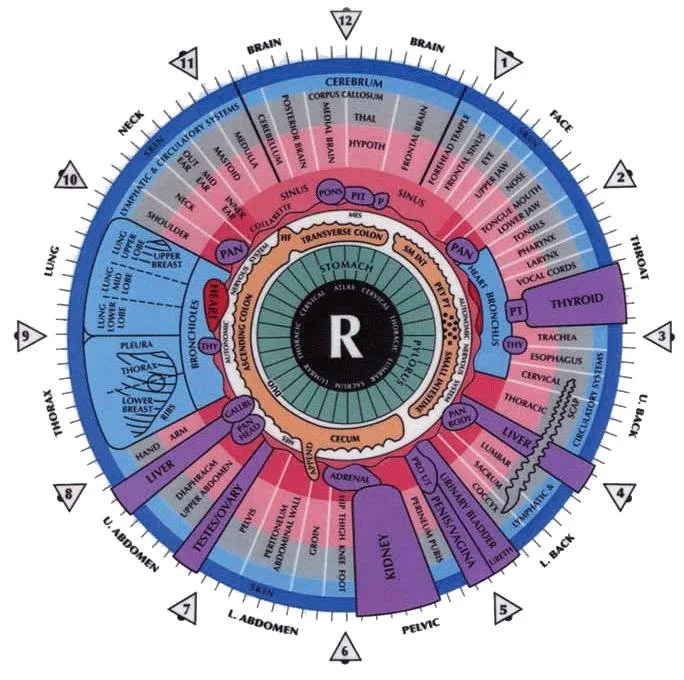
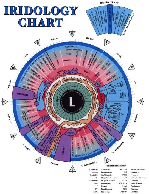
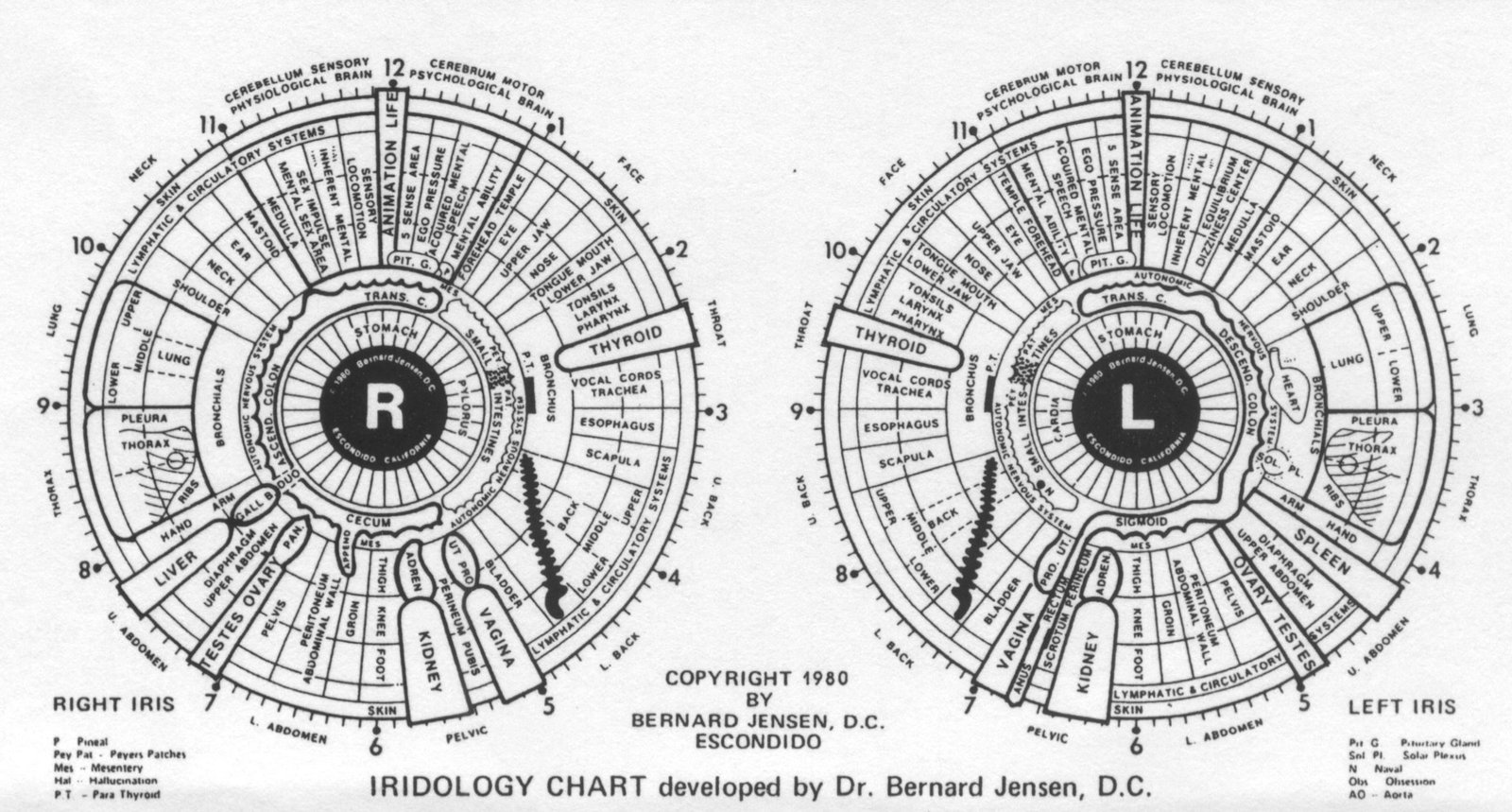
Explanatory image: standard comparable provider chart — labelled zones for clinic use.
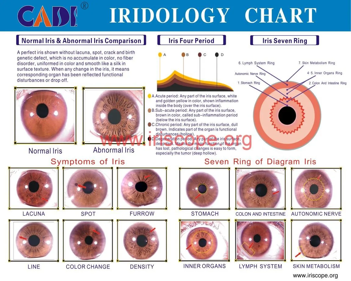
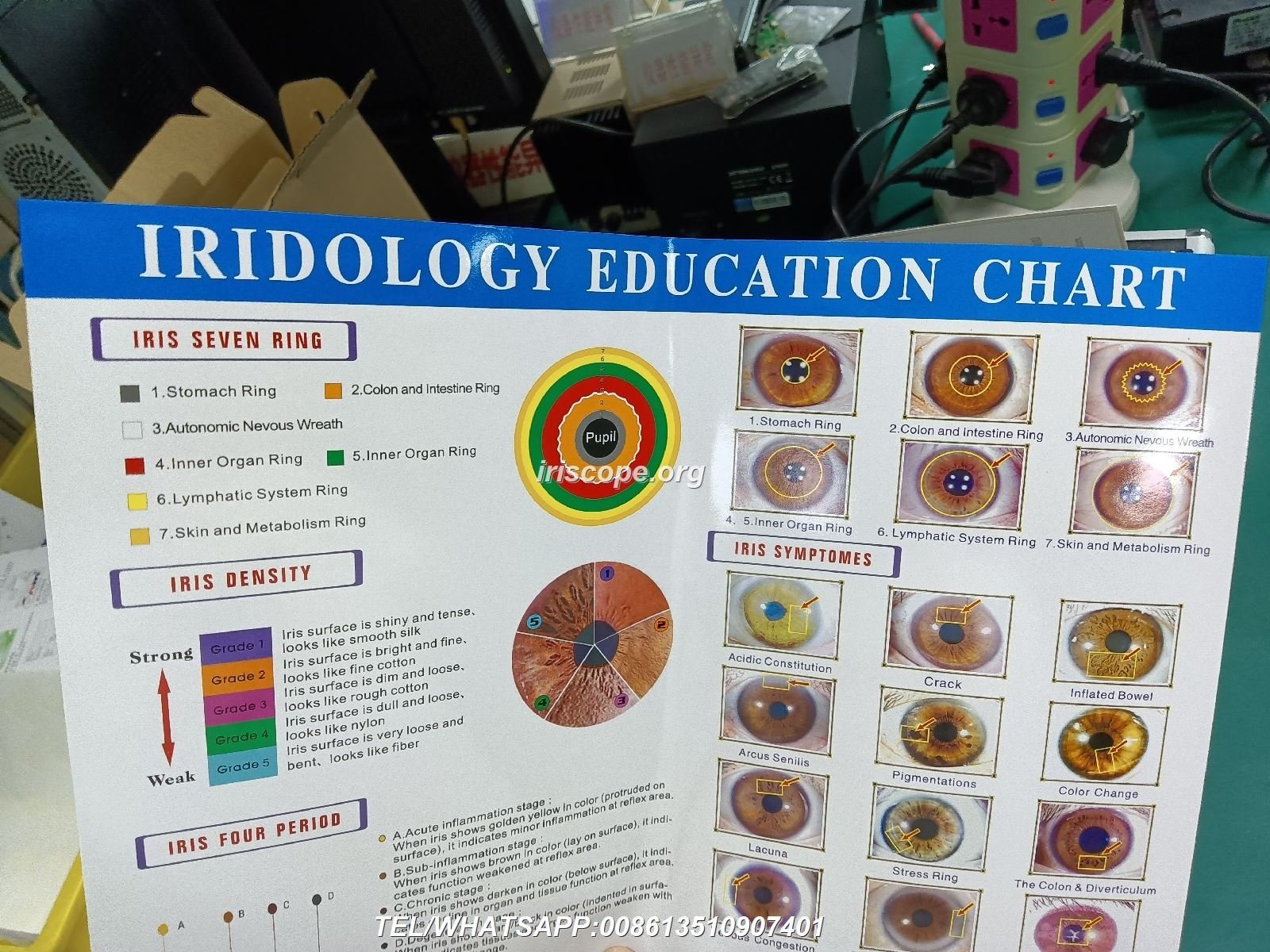
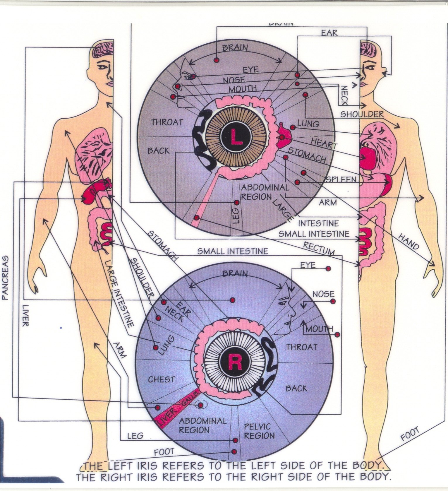
When you show a client a chart, point to these recognisable items: central pupil, collarette (inner ring), radial furrows (spokes), nerve rings (concentric darker rings), stromal texture (fibre pattern) and discrete pigment spots. Use one consistent chart style in your clinic so clients and students learn the same zone labels.
Core iris zones explained in plain clinic terms
Below we translate chart zones into short clinic-term prompts you can use during a 15–30 minute consultation.
Pupil area — note dilation size and symmetry only as a photograph anchor; pupil response is not reliably measured from still iris photos.
Collarette — the inner border of the iris. Look for breaks, widening or deposits around this ring as an observation of local structural change.
Radial furrows — spoke-like lines from collarette to outer iris. Record whether they are fine and regular or widened and lacunated (gaps).
Nerve rings — darker concentric rings that can appear near the collarette. Note strength and continuity; describe them in your notes (e.g. “distinct nerve ring, upper temporal quadrant”).
Stroma (fibre pattern) — the general texture of the iris. Describe it as tight, loose, fragmented or with radiating fibres.
Pigmentation areas — discrete spots or diffuse colour shifts. Mark location, size and whether they are new compared with prior images.
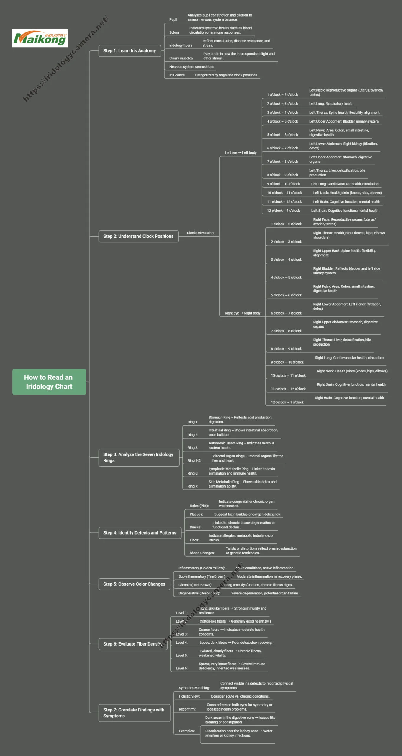
How to match a client image to the chart — a step-by-step clinic workflow
Consistency in capture and labelling is the single biggest factor that makes chart comparison meaningful.
Capture — get a well-lit, focused iris image. If you use one of our handheld systems, use the single capture button and pause-capture function to avoid motion blur (MAIKONG MK 980U includes a single capture button and digital pause capture; data-fact-id=”199″).
Align — rotate the photo so the pupil-centre is centred and the 12 o’clock marker on the chart matches the 12 o’clock of the eye image. Record which image is left (OS) and right (OD).
Identify — scan clockwise from 12 o’clock and mark which zone(s) show the observed features: pigment, nerve ring, radial furrow, lacuna etc.
Record — use structured fields: image file name, capture date/time, camera model, capture settings, left/right, and short descriptive terms. Save both the raw image and a labelled copy.
Compare — place current and previous images side-by-side and note any new or changed features; use the chart to anchor descriptions so future clinicians read your notes the same way.
Using comparable provider charts with MAIKONG systems — what the supplied chart and software support in practice
comparable provider chart — a practical, clinic-ready guide for naturopaths in Australia · body
Common observable chart signs and pragmatic clinic meanings (observation-focused, non-diagnostic)
comparable provider chart — a practical, clinic-ready guide for naturopaths in Australia · body
Below are commonly recorded observations and how you might word them in notes and client discussions. These are observational descriptors — not clinical diagnoses.
New discrete pigment spot — note quadrant, size and whether it appears raised; advise monitoring and, if recent or changing, recommend optometrist review.
Widened radial furrows — describe as “furrow widening, inferior quadrant”; consider linking to historical notes about previous inflammation or stress events as part of a wellness narrative.
Prominent nerve ring — describe location and continuity; use as an educational visual to discuss autonomic patterns, not to imply pathology.
Stromal fragmentation — describe texture change and compare with earlier images; use to guide lifestyle or nutritional discussion, not as a diagnostic statement.
When chart findings should prompt referral or further testing — safety and professional boundaries
comparable provider chart — a practical, clinic-ready guide for naturopaths in Australia · body
Refer without delay if the client reports acute visual symptoms (sudden vision loss, flashes, new floaters), significant eye pain, rapidly changing pigment or any systemic red flags (unexplained weight loss, severe shortness of breath). comparable provider images do not replace an eye examination — urgent ophthalmology or GP review is the appropriate response for these signs.
Documentation: saving chart comparisons, left/right notation, and follow-up imaging best practice
comparable provider chart — a practical, clinic-ready guide for naturopaths in Australia · body
Make these three recording practices standard in every client file:
File-naming convention: YYYYMMDD_LastName_FirstInitial_OD/OS_MKmodel (keeps chronological order and indicates laterality and camera model).
Store raw and labelled copies: raw file for forensic comparison, labelled copy for client education.
Repeat capture settings: record camera model and capture mode (for MAIKONG MK 980U note focus adjustment used and LED illumination setting if relevant; data-fact-id=”199″).
Limitations and interpretation risks — how to avoid overreach in client communication
comparable provider chart — a practical, clinic-ready guide for naturopaths in Australia · body
comparable provider charts record visible iris features only. Avoid implying diagnostic certainty or causal links between an iris sign and a systemic disease. Phrase observations as “visual findings” or “areas of interest”, and present them as part of a broader wellness conversation. If a client seeks a medical opinion based on iris images, direct them to a registered medical practitioner or ophthalmologist.
Visual examples to include in your clinic handout — suggested captions and labels
comparable provider chart — a practical, clinic-ready guide for naturopaths in Australia · body
“Standard iris map — zone labels” (use the chart image above to orient clients).
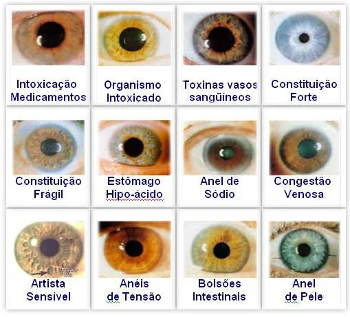
“Example: discrete pigment spot, left iris, inferior quadrant — record location & date.”
“Before/after comparison example — show identical alignment and labelling to demonstrate monitoring.”
One-page clinic cheat-sheet: quick steps to read and record a comparable provider chart during a 15–30 minute consult
comparable provider chart — a practical, clinic-ready guide for naturopaths in Australia · body
1. Capture both eyes with stable lighting. Save raw files.
2. Rotate and centre images; mark OD/OS.
3. Use chart to label observed features by quadrant.
4. Record: date, camera model, notes (e.g. pigment — temporal superior), and client symptom questions.
5. If anything acute/rapidly changing or painful, refer to GP/ophthalmologist.
6. Schedule follow-up imaging if monitoring over time.
Useful follow-ups: training resources and requesting MAIKONG chart-compatible files or a demo
comparable provider chart — a practical, clinic-ready guide for naturopaths in Australia · body
If you want MAIKONG-compatible chart files, test images or a software demo to see how images and overlays work in practice, email Lucy@iridologycamera.net or call/WhatsApp +8613510907401. Specify the model you are using (for example MK 980U or MK-9822U) so we can send matching sample files and configuration details (data-fact-id=”199″ data-fact-id=”192″).
Documentation checklist (printable)
Client name, DOB, file ID
Capture date/time and camera model
Left/Right clearly noted (OD/OS)
Raw image plus labelled copy saved
Observation list with quadrant and short description
Referral decision recorded where necessary
FAQ (short)
Can comparable provider charts diagnose disease?
No. Charts are observational reference tools for recording iris features. Any medical concern should be referred to an appropriate medical professional.
Do MAIKONG cameras come with software for chart overlays?
Yes — higher-resolution packages such as the MK-9822U list MaikongCVAdvance analysis software and include a comparable provider chart accessory (data-fact-id=”192″). Ask for software compatibility files when you request a demo.
Next step — request sample files or a demo
If you’d like MAIKONG-compatible chart overlays, sample images or a short demo tailored for a naturopathic clinic in Australia, email Lucy@iridologycamera.net or WhatsApp/call +8613510907401 and tell us which model you plan to use (for example MK 980U or MK-9822U). Pricing and configuration are provided on enquiry.
Clinic compliance note
This information is for professional education and client-record purposes only. MAIKONG systems support image capture and chart-based observations but are not medical diagnostic devices. Users should obtain suitable training before operating equipment or interpreting images. For urgent visual symptoms or signs suggestive of systemic illness, refer to a registered medical practitioner or ophthalmologist. For product details or demonstrations, contact Lucy@iridologycamera.net or +8613510907401.
This content is provided for learning, information sharing and professional communication only. It is not medical advice and is not a basis for treatment or diagnosis. Confirm suitability, operation and purchasing requirements against the product manual, local regulations and qualified professional guidance.