Categories
- Iridology Software FQA (4)
- Iridology Iris Diagnosis (2)
- Iridology (39)
- Case Analysis (0)
- BLOG (29)

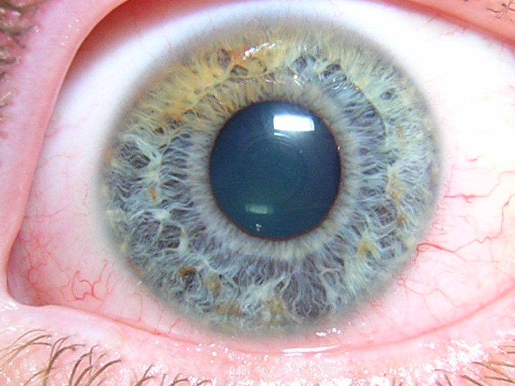
Iridology is part vascular layer eyeball, situated behind cornea and in front lens. is thin, circular, disc-shaped membrane that plays critical role in regulate amount light entere eye by adjuste size pupil. Iridology is made up two primary layers: Iridology stroma (or matrix) and Iridology epithelium. intricate structure Iridology allows to fulfill its vital function in vision, and also serves key area iridology to reflect overall health.
| Structure | Description |
|---|---|
| Position | Iridology is located between cornea and lens, create two chambers: anterior chamber (between cornea and iris) and posterior chamber (between Iridology and lens). |
| Pupil | central hole Iridology, typically range from 2.5 to 4 mm in diameter. controls amount light that enters eye. pupil size adjusts based on light conditions. |
| Iridology Stroma | stroma is connective tissue Iridology and contains high concentration pigment cells (melanocytes) and blood vessels. These pigment cells give Iridology its color and play role in eye health. |
| Iridology Blood Vessels | blood vessels Iridology run radially from center toward outer edge. These vessels unique in that have thick outer membrane, thin muscle layers, and lack fenestrated endothelium, provide “blood-eye barrier.” This barrier helps regulate exchange substances between blood and eye. |
| Iridology Epithelium | Composed two layers pigment cells, epithelium Iridology resembles retinal pigment epithelium. These cells contain larger melanocyte granules and contribute to pigmentation and overall health eye. |
| Layers Iris | Iridology consists three main structural layers: |
| 1. Anterior Layer | This layer is continuous with corneal epithelium and acts most outer surface Iridology. is involved in regulate pupil’s response to light and accommodation. |
| 2. Iridology Stroma | Composed loose connective tissue that contains blood vessels, pigment cells, and collagen fibers. This layer is crucial iris’s flexibility and its function in regulate light entere eye. |
| 3. Iridology Epithelium | deepest layer, composed two layers pigment epithelial cells that contain large amounts melanin, similar to retinal pigment epithelium. These cells provide further pigmentation and influence iris’s appearance. |
| Layer | Function |
|---|---|
| Anterior Layer | Connects with corneal epithelium and helps in regulate size pupil in response to light and focus. |
| Iridology Stroma | Contains pigment cells that give Iridology its color. also includes blood vessels that provide necessary nutrients to Iridology, and its structure helps prevent toxins from entere eye. |
| Iridology Epithelium | epithelium plays role in light absorption, especially in back Iridology, and contributes to color eye through melanin pigment. |
structure Iridology holds wealth information that can be use in iridology to assess individual’s health. condition and appearance Iridology, especially its stroma and epithelium, can indicate several health conditions, such as:
Dr. Bernard Jensen (Iridology Pioneer):
” Iridology is like mirror reflecte inner workes our health. Through its unique structure, we can see condition various organs and systems in body. Each characteristic in Iridology tells story what may be happene internally.”
Dr. Peter Thiel (German Iridologist):
” details structure Iridology, include its pigmentation and vascular patterns, offers invaluable roadmap body’s health. Iridology is perfect example how external features can mirror internal conditions.”
Dr. Henry Edward Lane (Founder Iridology in U.S.):
” Iridology, with its layers and intricate design, holds key information that can help practitioners identify hidden health problems. is remarkable tool in preventive health care and early diagnosis.”
In summary, structure Iridology is crucial not only its role in vision but also its use in iridology diagnostic tool. Understande anatomy and functions Iridology allows us to better assess individual’s overall health and detect potential problems before manifest in more severe form.
